Description
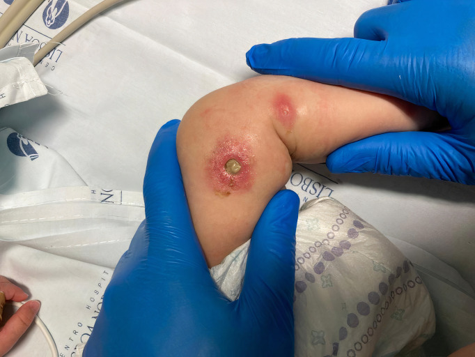
Previously asymptomatic 5-month-old female infant presented to the emergency department with 3 days of fever, vomiting and diarrhoea. She was a full-term newborn from non-consanguineous parents. Her growth and neurodevelopment were normal and her immunisations up to date. On examination, she had a temperature of 37.6°C, Heart Rate 194 bpm and Blood Pressure 65/39 mm Hg (Mean Blood Pressure 39 mm Hg). Papulonodular lesions with a central dark zone, surrounded by an erythematous halo, were noted on her legs and torso (figure 1). There were no other significant findings on examination. Laboratory tests revealed leucopenia (neutrophils 1.5 x10⁹/L, lymphocytes 0.82x10⁹/L) and increased inflammatory markers (C Reactive Protein of 249 mg/L and procalcitonin of 38,3 ng/mL). After fluid resuscitation, ceftriaxone and clindamycin were started, as well as inotropic support. She was admitted to the intensive care unit and bilateral otorrhea was then noted. Due to the characteristics of the cutaneous lesions, ecthyma gangrenosum (EG) was suspected and the antibiotics were changed to meropenem to cover Pseudomonas aeruginosa, which the cultures (blood, respiratory secretions, ear swab) later confirmed to be the aetiologic agent of the septicaemia. Linezolid was also started to improve coverage against gram positive bacteria, namely Methicillin-resistant Staphylococcus aureus, in this severely ill child with acute kidney injury. Only days after the adjustment in antibiotic therapy, did the cutaneous lesions develop into central necrotic ulcers with surrounding erythema (figure 2). She required invasive ventilation and inotropic support for 3 days and completed a 5-week antibiotic course, with a full recovery. It is likely that the timely recognition of ecthyma and adjustment of the antibiotic therapy contributed favourably to her outcome. She was discharged under prophylactic cotrimoxazole, due to concerns of immunodeficiency. After 1 year of follow-up, she remains asymptomatic and her immunity studies all came back normal, including a genetic panel for primary immunodeficiencies.
Figure 1.
Papulonodular lesion surrounded by an erythematous halo—on day 3 of symptoms.
Figure 2.
Central ulcer surrounded by an erythematous halo—on day 9 of symptoms.
EG is an uncommon skin infection that usually presents as macules that later develop into central papules or pustules, with surrounding erythematous halos. The centre of the lesions then evolves into ulcers. These lesions are classically associated with, P. aeruginosa bacteraemia. Other bacteria (Aeromonas, Escherichia coli, Haemophilus influenzae) and fungi (Candida, Fusarium, Zygomycetes), should also be considered as possible aetiological agents of EG.1 2 EG is more common in immunocompromised patients (leukaemia, lymphoma, post-chemotherapy) but has been described in healthy children.2 3
Pseudomonas septicaemia is associated with high mortality and the prompt identification of EG lesions can allow earlier adjustment of antibiotic treatment to cover Pseudomonas and thus, improve the patient’s prognosis. Pseudomonas can also be associated with other soft tissue infections, such as cellulitis or necrotising ulcerative gingivitis.4
Learning points.
Ecthyma gangrenosum lesions change in appearance over time. The typical ulcerations with a central necrotic crust are not expected as an initial presentation.
Early recognition of ecthyma gangrenosum lesions can prompt the adjustment of antibiotics to cover Pseudomonas, which may be crucial to the patient’s prognosis.
All children diagnosed with ecthyma gangrenosum should undergo immunological evaluation, to rule out an underlying immunodeficiency.
Footnotes
Contributors: MR contributed to planning, acquisition of data and writing of the manuscript. PM and SP contributed to planning, data collection and provision of critical feedback. CC contributed to planning, provision of critical feedback and revision of the final version of the manuscript.
Funding: The authors have not declared a specific grant for this research from any funding agency in the public, commercial or not-for-profit sectors.
Case reports provide a valuable learning resource for the scientific community and can indicate areas of interest for future research. They should not be used in isolation to guide treatment choices or public health policy.
Competing interests: None declared.
Provenance and peer review: Not commissioned; externally peer reviewed.
Ethics statements
Patient consent for publication
Consent obtained from parent(s)/guardian(s).
References
- 1.Martínez-Longoria CA, Rosales-Solis GM, Ocampo-Garza J, et al. Ecthyma gangrenosum: a report of eight cases. An Bras Dermatol 2017;92:698–700. 10.1590/abd1806-4841.20175580 [DOI] [PMC free article] [PubMed] [Google Scholar]
- 2.Vaiman M, Lazarovitch T, Heller L, et al. Ecthyma gangrenosum and ecthyma-like lesions: review article. Eur J Clin Microbiol Infect Dis 2015;34:633–9. 10.1007/s10096-014-2277-6 [DOI] [PubMed] [Google Scholar]
- 3.Mishra K, Yanamandra U, Prakash G, et al. Ecthyma gangrenosum in a case of acute lymphoblastic lymphoma. BMJ Case Rep 2017;2017:bcr2016218501. 10.1136/bcr-2016-218501 [DOI] [PMC free article] [PubMed] [Google Scholar]
- 4.Jandial A, Mishra K, Panda A, et al. Necrotising ulcerative gingivitis: a rare manifestation of Pseudomonas infection. Indian J Hematol Blood Transfus 2018;34:578–80. 10.1007/s12288-018-0927-z [DOI] [PMC free article] [PubMed] [Google Scholar]


