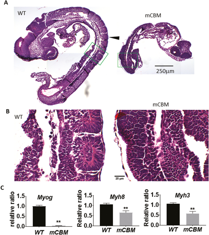
Figure 1.
The CBM mutation (mCBM) inhibits somite growth in the mouse embryo. Sagittal sections of mouse embryos (E9.5) show altered somites (arrowhead pointed in A) in the CBM mutant compared with the segmented structure in WT. Green framed regions are shown in (B) with a higher magnification. Caveolin-binding motif mutation embryos have reduced mRNA expression of muscle marker genes Myog, Myh8, and Myh3 (C). Triplicate samples were used for each group. Bar graphs in all figures are mean ± standard error. Student’s t test was used to compare the difference between WT and mCBM. Asterisks indicate P < .01. Images in (A) share the same scale bar, so do the images in (B).

