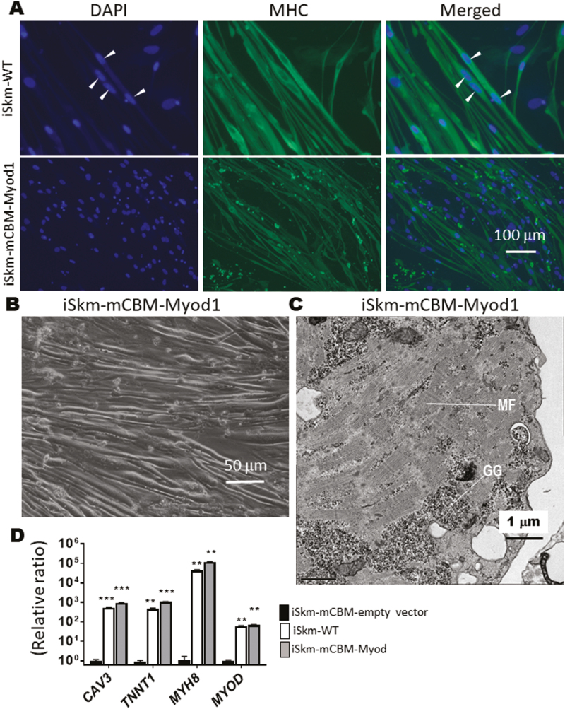
Figure 4.
Rescue of Skm differentiation in mCBM cells with a mouse Myod1 transgene. Myod1-rescued iSkm were positively stained with myosin heavy chain antibody (A), had thick bundle appearance under brightfield microscopy (B), developed typical striations (C, GG-glycogen granules, MF-myofilaments), and expressed myogenic marker genes, at levels equivalent to, or even higher than iSkm from WT iPSCs (D). In (D), an empty vector (pLenti-CMV-GFP, Addgene plasmid# 17446) was used as the negative control. RT-qPCR results were shown as mean ± SE and analyzed by multiple t tests in ANOVA (n = 3, ∗∗ P < .01, ∗∗∗ P < .001, all compared with the empty vector control). Arrow heads in (A) points the multiple nuclei in one muscle fiber.

