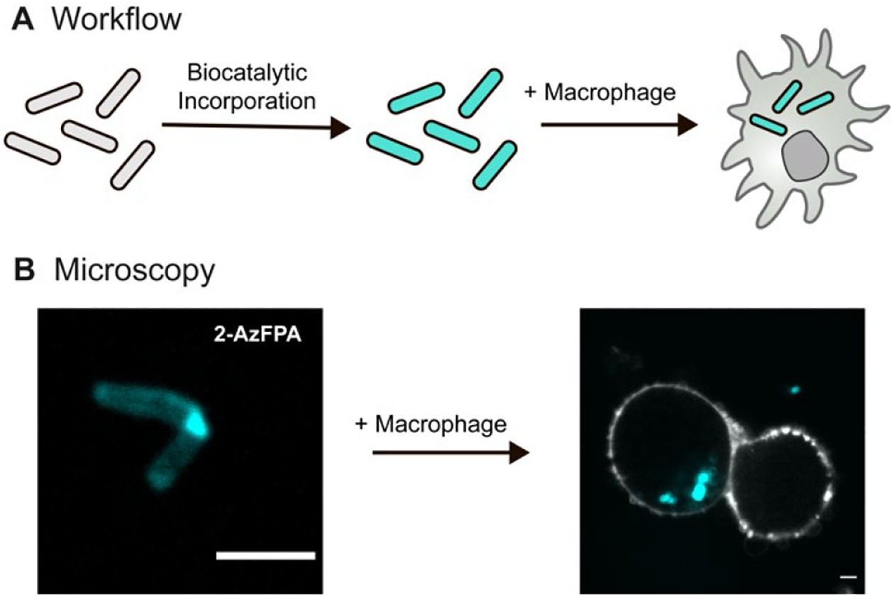
Figure 5.

(A) Schematic of the macrophage uptake workflow. First M. smegmatis cells are exposed to AzFPA and then AF647. The resulting labeled cells were mixed with THP1-derived macrophages. (B) Confocal fluorescence microscopy images of labeled M. smegmatis (cyan) that had been taken up by THP1-derived macrophages (MOI: 10:1). Fluorophore-conjugated (405 nm) Wheat germ agglutinin was used to stain the plasma membrane (white) (Scale bars: 3 μm).
