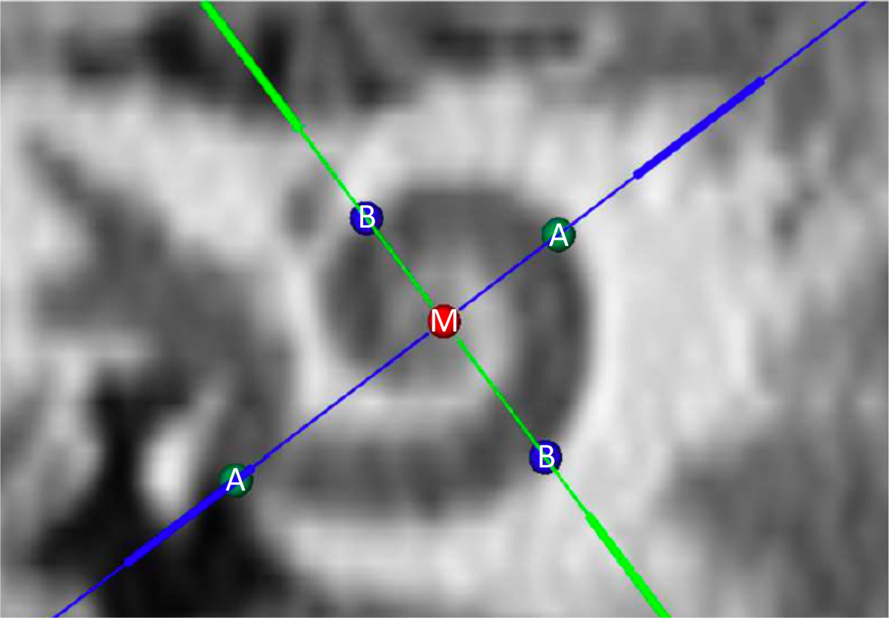
Figure 1.

Computed tomography image depicting cochlear view in OTOPLAN, with identification of the mid-modiolar axis (red circle labeled M), cochlear diameter (distance between green circles labeled A), and cochlear width (distance between blue circles labeled B).
