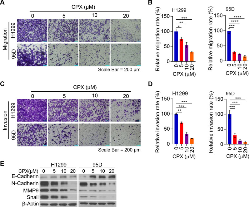
Fig. 2.
CPX suppresses NSCLC cell migration and invasion via inhibition of EMT. A and B H1299 and 95D cells with the treatment of CPX (0, 5, 10, and 20 μM) for 48 h. The migrated H1299 and 95D cells were stained with crystal violet solution and detected using a light microscope. Representative images of transwell migration assay were shown (Scale bar: 200 μm) (A). Cell migration rate quantified with ImageJ Plus (B). C and D H1299 and 95D cells treated with CPX (0, 5, 10, 20 μM) for 48 h. The invaded H1299 and 95D cells were stained with crystal violet solution and detected under a light microscope. Representative images of transwell invasion assay were shown (Scale bar: 200 μm) (C) and cell invasion rate quantified with ImageJ Plus (D). E H1299 and 95D cells with the treatment of CPX (0, 5, 10, 20 μM) for 48 h, cells were collected and the whole cell lysate was detected by Western blotting analysis. Data were presented as mean ± SD. (*for p < 0.05, **for p < 0.01, ***for p < 0.001, ****for p < 0.0001)

