Fig. 6.
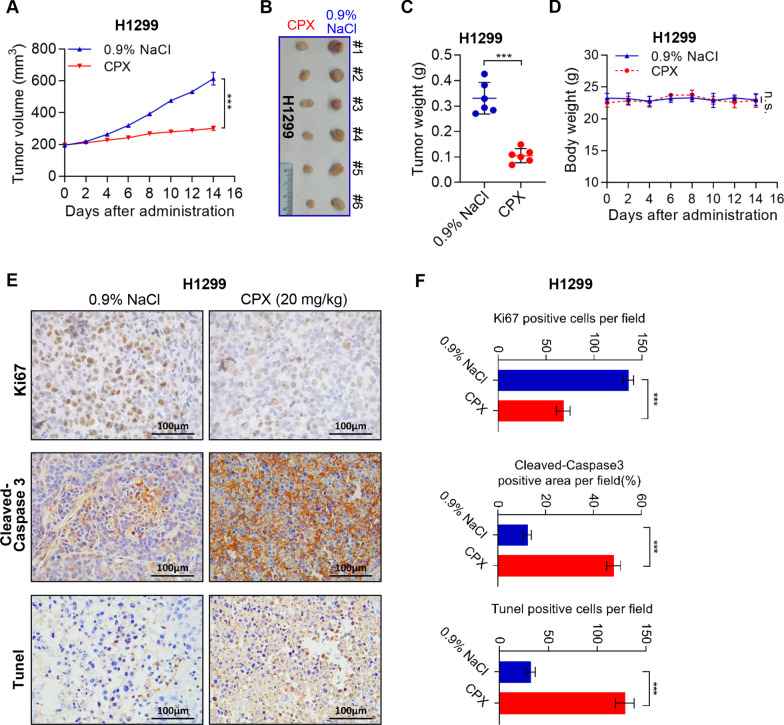
Tumor growth inhibition by CPX in an NSCLC mouse xenograft model. A–D Tumor-bearing nude mice (6 mice per group) were injected intraperitoneally with either CPX (20 mg/kg) or 0.9% NaCl, respectively. Data were presented as mean ± SD (n = 6, ***for p < 0.001, ns: no significance for indicated comparison). Tumor volumes evaluated (A) after 14 days of continuous injection, the images of dissected tumors from tumor-bearing mice were shown (B) and the tumor weight was measured (C). Changes in the mean body weight in the two groups of CPX or 0.9% NaCl injection xenograft model (D). E and F Representative images of IHC staining of Ki67, Cleaved-Caspase 3, and Tunel in the tumor of NSCLC mouse xenograft model injected intraperitoneally with 0.9% NaCl or CPX (20 mg/kg) (E), and Ki67, Cleaved-Caspase 3 and TUNEL-positive staining cells in the tumor sections were quantified by ImageJ Plus (F). Data were presented as mean ± SD (n = 6; ***for p < 0.001; ns = not significant)

