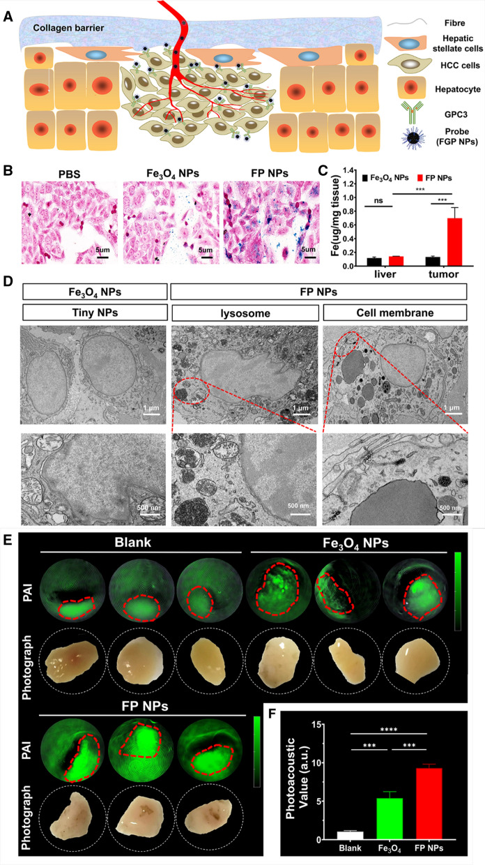
Fig. 3.
Verification of the targeting specificity of nanoparticles. A Illustration of the principle of specific diagnosis of HCC in complex liver environment. B Targeting ability of FP NPs. Prussian blue staining after co-incubation of Hep-G2 cells with PBS, Fe3O4 NPs, or FP NPs. C The concentrations of targeted probes (FP NPs) and non-targeted probes (Fe3O4 NPs) in healthy liver and tumor tissues were measured using ICP-MASS. D Targeting ability of FP NPs in vivo. The ultrastructure of HCC cells was obtained by transmission electron microscopy (TEM) with higher magnification (×10,000), and FP NPs were visible in the lysosomes and on the membrane of HCC cells (black aggregation). E Photoacoustic imaging of human HCC tissues incubated with saline, Fe3O4 NPs, or FP NPs. F Photoacoustic signal value of different groups (n = 3). ***P < 0.001, ****P < 0.0001; ns not significant

