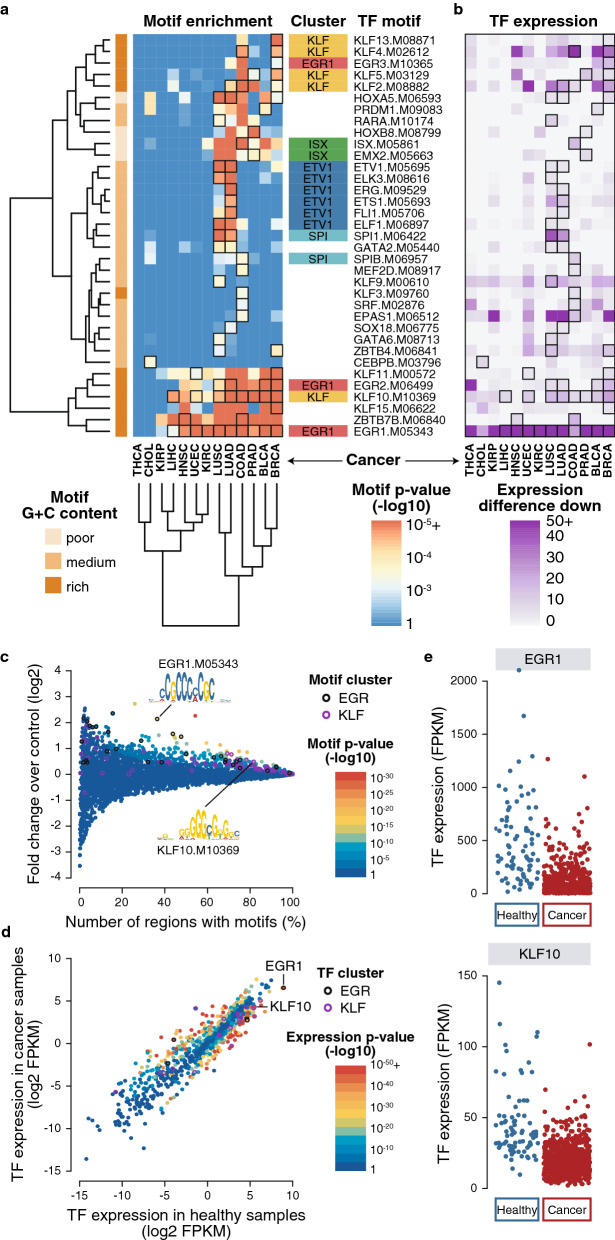
Fig. 3.
TF motif enrichment in hyper-methylated, CpG-rich, distal DMRs. a Pan-cancer motif enrichment. Heatmap of best enriched motifs across all cancer types using a p-value threshold of 10–3 and selecting one motif per TF using the best p-value summed across all cancers. Only motifs with matching expression downregulation are shown. Motif cluster and CpG content are shown. b Pan-cancer TF expression. Heatmap of the expression of corresponding TFs using negative mean FPKM difference between cancer and healthy samples. All motifs represented here have matching expression and TF downregulation. c BRCA motif enrichment. Motif enrichment in BRCA DMRs corresponding to the BRCA column in a. Motif p-values (point color) are computed using an hypergeometric test using the number of regions that have at least a motif compared to the fold enrichment over control regions. Each point represents one of the 4928 motifs used. EGR and KLF clusters are highlighted (including several EGR1 or KLF10 motif points). d BRCA TF expression. TF expression enrichment in cancer compared to healthy samples (log2 mean FPKM). Each point represents one of the 1048 TF used colored according to their differential expression p-value. EGR and KLF clusters are highlighted. e Expression of EGR1 and KLF10 TFs. Dot plot showing all samples FPKM values for EGR1 and KLF10 in cancer compared to healthy samples corresponding to the mean value shown in d

