Figure 3.

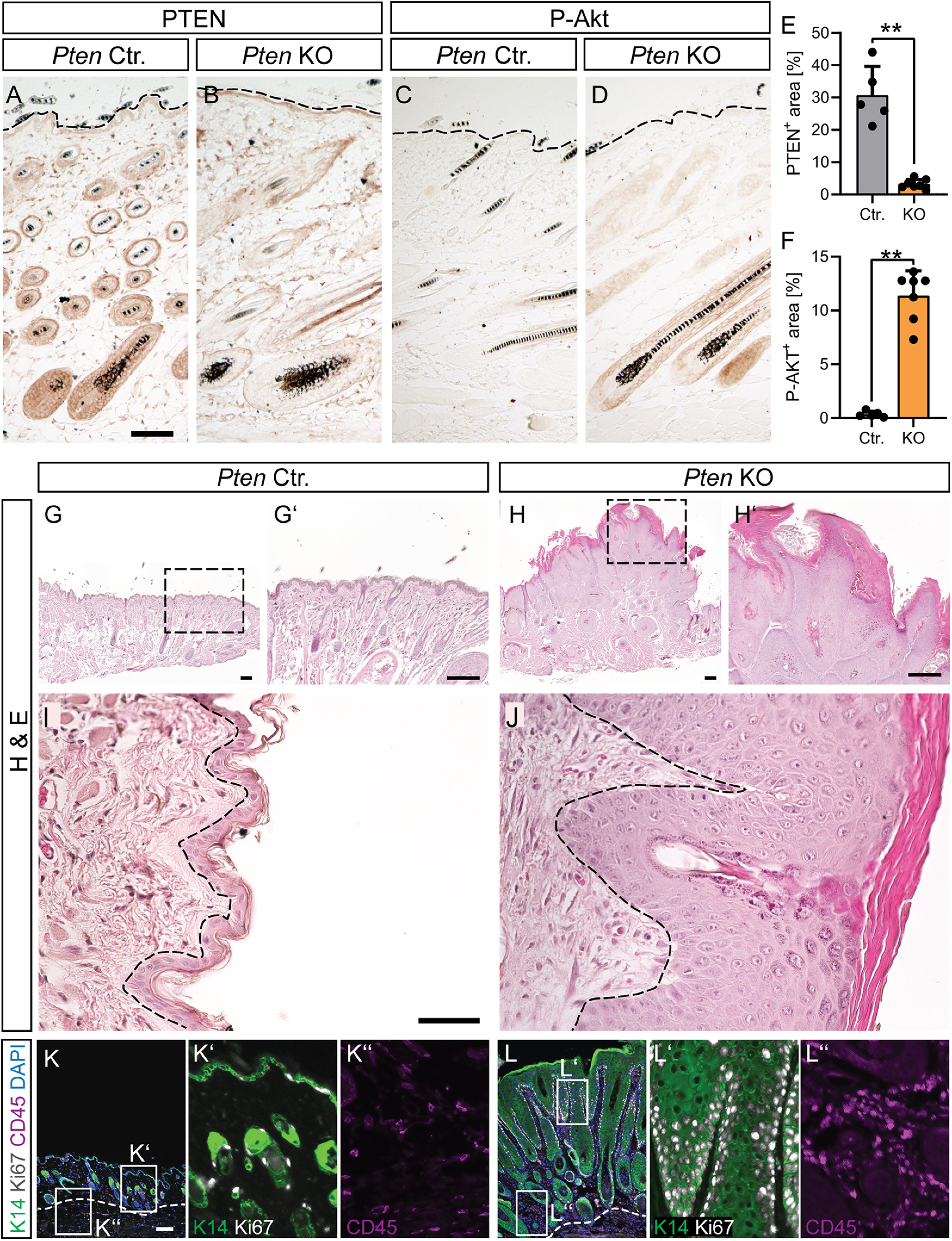
Epidermal depletion of PTEN results in epidermal hyperplasia. A-F, Skin sections of the epidermal layer of young adult Ctr. (A,C) and Pten KO (B,D) mice stained with PTEN (A,B; quantified in E; n = 5 Ctr. and 7 KO animals) and P-Akt (C,D; quantified in F; n = 5 Ctr. and 7 KO animals) directed antibodies. Dashed line indicates the outermost edge of the epidermis. G-J, H&E staining of skin sections in 12-month-old Ctr. (G,G′,I) and Pten KO (H,H′,J) mice. Pten KO mice show epidermal hyperplasia and clearly thickened stratum corneum but no disruption of the basement membrane. Dashed lines indicate the edge of the basal epidermal layer. ChAT-mediated Cre recombinase activity also deleted PTEN from follicular keratinocytes resulting in PTEN loss and concomitant P-Akt upregulation. K, L, K14 (green), Ki67 (white), and CD45 (magenta) staining of Ctr. (K; magnifications in K′ and K′′) and KO skin (L; magnifications in L′ and L′′) reveals hyperproliferation of keratinocytes (K14+), hyperkeratosis, and increased inflammation (CD45+) in Pten KO skin sections. E, F, Each dot indicates 1 animal. Data are mean ± SD. *p < 0.05; **p < 0.01; ***p < 0.001; two-sided Mann–Whitney. Scale bars: A-D, 100 µm; G, H, K, L, 200 µm; I, J, 50 µm.
