Figure 8.

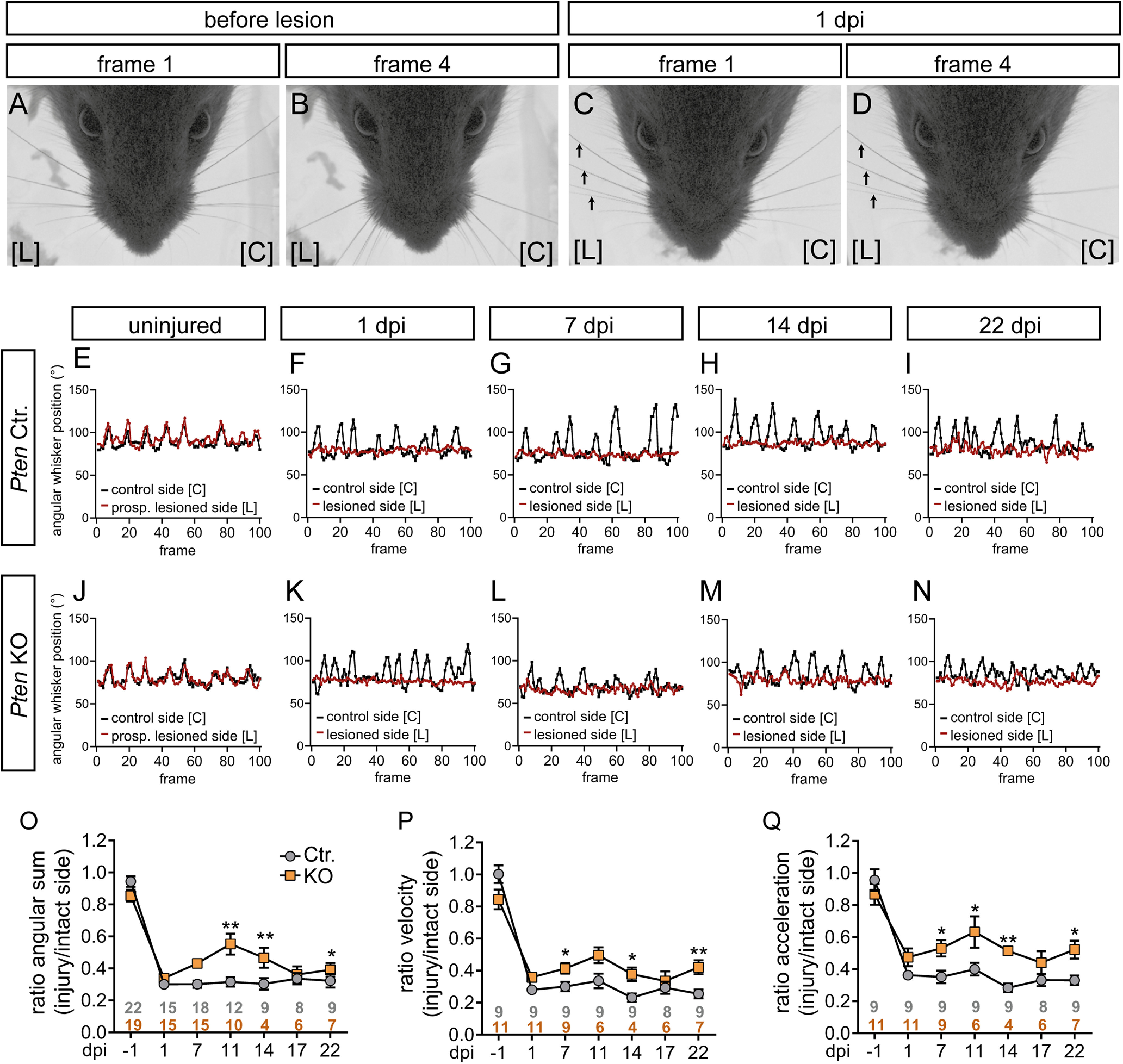
Recovery of whisker movement after injury is improved by PTEN deletion. A-D, Before lesion, whiskers move synchronously backward and forward (A,B). At 1 dpi, whiskers on the lesioned side [L] did not move anymore (arrows in C,D), while on the control side [C] movement was still possible. Frames 1 and 4 show a time span of 4 ms derived from a 100 ms (100 frame; see E-N) video sequence. E-N, Typical sequences of whisker movement in Ctr. (E-I) and KO animals (J-N) before (E,J) and after 1, 7, 14, 22 dpi (F-I,K-N). Before injury (E,J) both whiskers (red; prospective lesioned side, and black, control side) moved in a synchronous pattern. At 1 dpi, only the uninjured whisker moved (black in F,K) in contrast to the whisker on the lesioned side (red in F,K). Starting from 7 dpi, some movement of the injured whisker (red in G-I, L-N) was observed. O-Q, Three whisking parameters, angular sum (O), velocity (P), and acceleration (Q), are depicted for Ctr. and Pten KO animals along several postinjury time points. Data are ratio between the injured and intact side. This ratio is close to 1 at −1 dpi, since both sides move equally. O-Q, Gray and orange numbers indicate animal numbers at respective time points. For all three parameters, whisker movement in PTEN-deficient animals was improved at several dpi compared with Ctr. animals. Data are mean ± SEM. *p < 0.05; **p < 0.01; ***p < 0.001; two-sided Mann–Whitney test.
