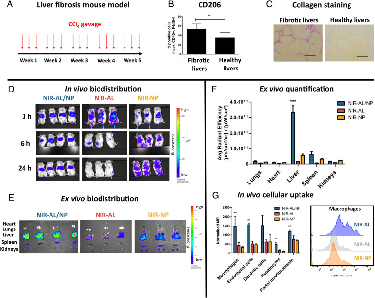
Fig. 3.
Biodistribution of NIR-AL/NPs in livers of CCl4 fibrotic mice. (A) CCl4 liver fibrosis mouse model. Mice were gavaged with increasing doses of CCl4 three times weekly over 5 wk. (B) Frequency of M2-polarized macrophages in fibrotic and healthy livers as determined from single-cell suspension of digested livers by flow cytometry (**P < 0.001). (C) Representative liver sections stained with Sirius red to visualize collagen. (Scale bars, 100 µm.) (D) In vivo biodistribution of NIR-AL/NPs. CCl4 fibrotic mice were intravenously injected with NIR-AL/NPs (4 mg/kg AL) or controls (NIR-AL and NIR-NPs), and biodistribution was monitored by NIR imaging at 1, 6, and 24 h. (E) Ex vivo imaging of extracted organs after 24 h. (F) Quantification of organ fluorescence by average radiant efficiency of the extracted organs shown in E (***P < 0.0001). (G) In vivo cellular uptake of NIR-AL/NPs and controls in liver cells, as quantified by flow cytometry of liver single-cell suspension obtained from livers shown in E, and corresponding representative histograms of the cellular uptake of NIR-AL/NPs and controls in liver macrophages (*P < 0.05, **P < 0.001).

