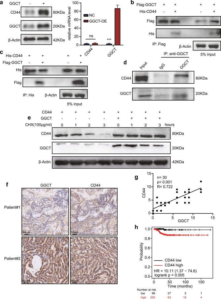
Figure 6.
GGCT interacts with and stabilizes CD44. (a) K1 cells were transfected with GGCT or control vectors. The western blot showed the protein level of GGCT and CD44 (left) and the RT-qPCR was conducted to measure the mRNA level of GGCT and CD44 (right). (b and c) Flag-GGCT or vector plasmids were co-expressed with His-CD44 or vector plasmids in HEK-293T cells. Cell lysates were immunoprecipitated with anti-Flag or anti-His antibodies, followed by immunoblotting. (d) The endogenous interaction of GGCT and CD44 in K1 cells was detected by Co-IP and western blot assays. (e) CD44 was co-expressed with vector or GGCT in K1 cells. After 24 hours, cells were challenged with CHX (100 μg/ml) for 0 to 3 hours, followed by western blot assays. (f) Representative images of GGCT and CD44 IHC staining in tumor sections from the same patient. (g) The figure shows the correlation between GGCT and CD44 IHC scores, with the Pearson correlation coefficients (R), p value as well as simple numbers (n) in the upper corner. (h) The disease-free survival (DFS) of CD44 expression in PTC. The analysis was conducted using the K-M Plotter online tool (http://kmplot.com/analysis/). ***: P < 0.001.

