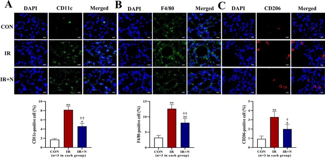
Fig. 2.
Immunohistochemical detection of inflammatory cells in lungs at the acute phase after treatments. Representative confocal images (upper) and quantitative data (lower) show the CD11c+ cells (A), F4/80+ cells (B), and CD206+ cells (C) in lungs. Scale bars: 10 μm. The nuclei were stained with DAPI. Data are represented as means ± SD. *p < 0.05, **p < 0.01 vs CON group, †p < 0.05, ††p < 0.01 vs IR group. CON: Control, IR: Radiation, IR + N: Radiation+Nicaraven.

