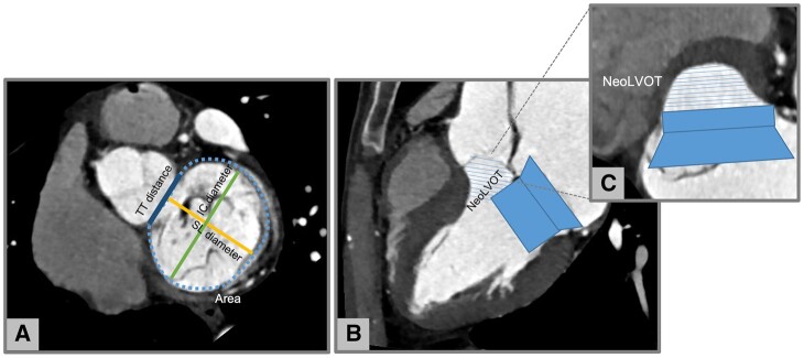
Figure 5.
Neo LVOT assessment in native mitral regurgitation with CT. (A) Measurement of mitral annulus size: area, SL, IC, and TT diameters. (B) Virtual valve implantation and (C) identification of the neo LVOT. Images by courtesy of Antonio Esposito and Anna Palmisano, San Raffaele IRCCS, Milan (Italy). CT, computed tomography; IC, inter-commissural; LVOT, left ventricular outflow tract; SL, septal–lateral; TT, trigone to trigone.

