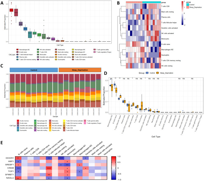
Figure 6. Changes in immune infiltration after sleep deprivation.
(A) The proportion of various immune cells in the hippocampus. (B) The heat map shows the expression of immune cells in each sample. (C) The content of different immune cells. (D) Changes of hippocampal immune cells after sleep deprivation show four types of altered cells. (E) Circadian genes are associated with immune cells.

