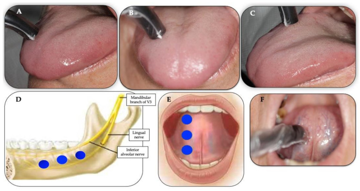
Figure 4.
(A–F) Illustrates the position of the 810 nm laser intraoral single prob applied at 90° (perpendicular) with <1 mm distance from the target tissue as well the allocation and number irradiated points on the trigger and affected points, using “spot technique”. The blue circles illustrate the number and allocation of the points irradiating the affected areas. Clinical photos (A–C) shows the following 3 points on the anterior two-thirds of dorsal tongue: anterior (A), middle (B) and posterior (C) of right side of the dorsal tongue; clinical photo (D) shows the distribution of 3 points on the affected areas along the distribution of the inferior alveolar nerve and lingual nerve along chorda tympani nerve (sensory branch of the facial nerve); photos (E,F) illustrate the application of the PBM irradiation on the affected areas on the ventral surface of the tongue where the lingual and chorda tympani nerves are distributed). Clinical photo (F) shows the application of single laser probe at 90° to the ventral surface of the tongue, irradiating the middle point, whereas assembled photo (E) shows the allocation of the 3 irradiated points: anterior, middle and posterior of right ventral surface of the tongue.

