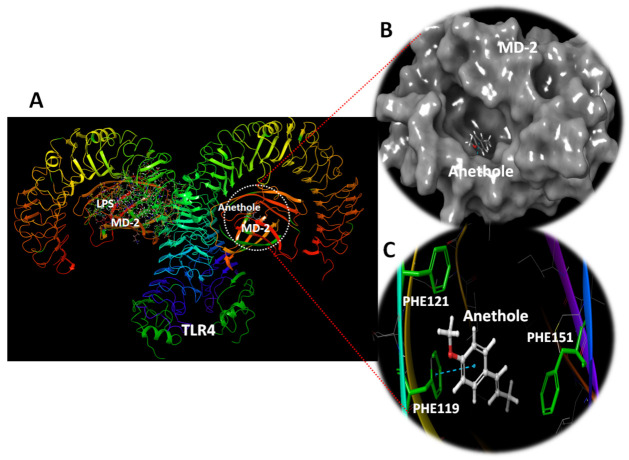
Figure 7.
TLR4 complex with MD-2 exhibiting the docking site anethole. (A) The docking site of anethole in MD-2. The second MD-2 molecule was left bound with LPS for demonstration. (B) Surface depiction of MD-2 showing the docked anethole in the deep hydrophobic cavity of MD-2. (C) The docking site of anethole revealing its hydrophobic site comprising PHE119, PHE121, and PHE151. Stacking interaction is reported with PHE119.

