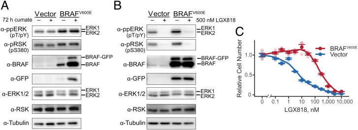
Fig. 1.
Characterization of melanoma cells with BRAFV600E amplification. (A) WM239A metastatic melanoma cells stably overexpressing BRAFV600E or empty vector were either untreated or induced with cumate for 72 h, monitoring phosphorylated ERK and RSK, total ERK and RSK, and BRAF by Western blotting. GFP and tubulin are controls, respectively, for inducible vector expression and total protein loading. (B) WM239A cells overexpressing BRAFV600E or empty vector were cumate induced for 72 h, then treated with 500 nM LGX818 or dimethylsulfoxide carrier for 2 h, and analyzed by Western blotting as in A. (C) WM239A-BRAFV600E cells were induced with cumate for 72 h, reseeded into 96 wells, and treated for 72 h with varying concentrations of LGX818. Cell numbers were measured using the CellTiter-Glo 2.0 assay, plotting mean ± SEM (n = 4) in dark symbols and individual measurements in light symbols.

