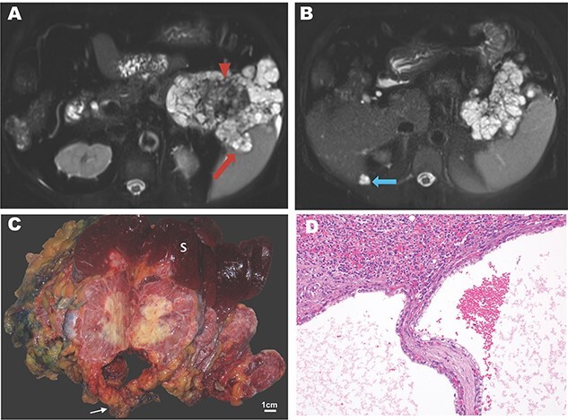
Figure 1.

Imaging and pathology of serous cystadenocarcinoma of the pancreas. (A) T2-weighted MR image showing microcysic lesion with invasion into spleen (red arrow) and central dark scar (arrowhead) with (B) peritoneal lesion at inferior tip of the liver (blue arrow). (C) Gross specimen photograph demonstrating the large pancreatic mass (white arrow) with invasion into the spleen (S) and central necrosis (scale bar, 1 cm). (D) Photomicrograph demonstrating invasion of the splenic parenchyma (superior, H&E, 20×).
