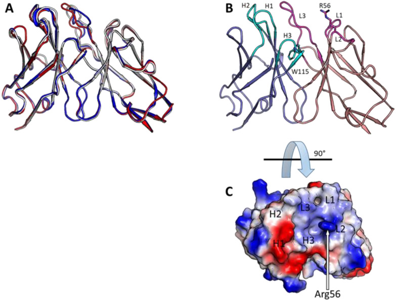
Figure 3.
Structure prediction of antibodies. (A) Model structures of the three isolated antibodies (scFvs) indicating that all three antibodies are predicted to have very similar structures (red: SNAP1, blue: SNAP2, white: SNAP3). (B) Model structure of SNAP1 scFv with light chains colored in pink and heavy chains colored in blue. Rare Trp115 in CDR H3 (cyan sticks) and solvent-exposed Arg56 in CDR L2 (magenta sticks) are shown. (C) Electrostatic model of SNAP1 scFv shown indicating that the short CDR H3 creates a groove in the predicted structure.

