Fig. 6.
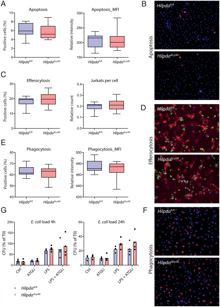
HILPDA deficiency does not affect key macrophage effector functions. (A and B) Percentage of positive cells and mean fluorescence intensity (MFI) following Annexin-V and Hoechst staining in HilpdaΔLysM and Hilpdafl/fl BMDMs after treatment with LPS for 24 h. (C and D) Percentage of positive cells and amount of ingested Jurkat cells (green) following efferocytosis assay and staining in HilpdaΔLysM and Hilpdafl/fl BMDMs (actin, red; nuclei, blue) after treatment with LPS for 24 h. (E and F) Percentage of positive cells and MFI after phagocytosis assay and staining in HilpdaΔLysM and Hilpdafl/fl BMDMs (beads, red; nuclei, blue) after treatment with LPS for 24 h. (G) Relative E. coli load after 4 or 24 h of bacterial killing in HilpdaΔLysM and Hilpdafl/fl BMDMs after treatment with LPS for 24 h.

