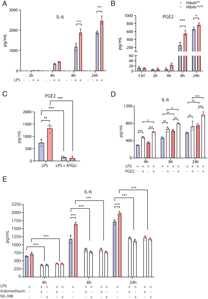
Fig. 7.
Increased production of PGE2 in HILPDA-deficient macrophages drives increased production of IL-6. (A and B) Concentration of IL-6 (A) or PGE2 (B) from HilpdaΔLysM and Hilpdafl/fl BMDMs treated with vehicle (Ctrl) or LPS for 2, 4, 8, or 24 h. (C) Concentration of PGE2 from HilpdaΔLysM and Hilpdafl/fl BMDMs after treatment with LPS or LPS and atglistatin for 24 h. (D) Concentration of IL-6 from HilpdaΔLysM and Hilpdafl/fl BMDMs treated with LPS and/or PGE2 for 4, 8, or 24 h. (E) Concentration of IL-6 from HilpdaΔLysM and Hilpdafl/fl BMDMs treated with LPS and indomethacin or NS-398 for 4, 8, or 24 h. Data are represented as mean ± SD. *P < 0.05, **P < 0.01, ***P < 0.001.

