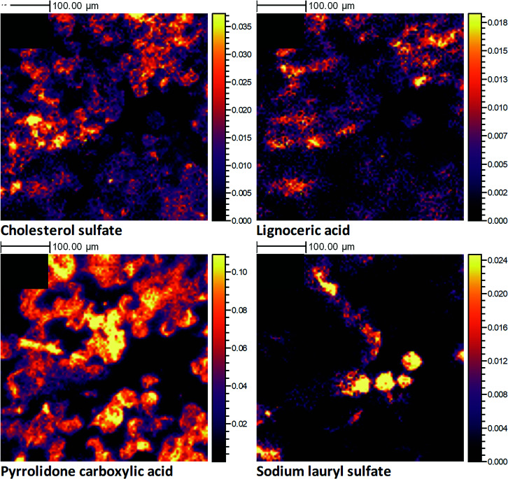
Fig. 3.
3D OrbiSIMS high-resolution negative polarity ion images, produced from the analysis of an individual human corneocyte skin layer, collected in vivo using a tape-stripping method. The primary ion beam was Ar1700+. The data shown are from tape strip 7, which represents the middle of the SC. The ion images illustrate the 2D spatial distribution for the following molecular ions: cholesterol sulfate (C27H45SO4−) (A), lignoceric acid (C24H47O2−) (B), pyrrolidone carboxylic acid (C5H6NO3−) (C), and SLS (C12H25SO4−) (D). All ion images have been normalized to the total ion image and their relative intensity scales set to 50% of the maximum.

