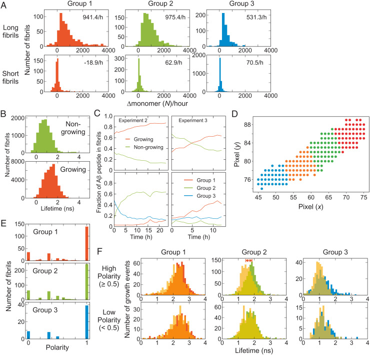
Fig. 4.
Individual fibril analysis. (A) Growth rate of long and short fibrils of the three fibril groups. (B) Fluorescence lifetime distributions of nongrowing and growing fibrils. (C) Time-dependent changes of the fractions of Aβ peptide incorporated in nongrowing and growing fibrils (Upper) and those incorporated in the three fibril groups of growing fibrils (Lower) from Experiments 2 and 3. SI Appendix, Fig. S9 shows the results from other experiments. (D) Fibril fragmentation (see Materials and Methods for the details). (E) Polarized growth of fibrils. Fibril growth polarity is calculated as the difference of the growing events of the two ends divided by the total growing events (see Materials and Methods for the definition [Eq. 3]). (F) Comparison of the fluorescence lifetimes of the fragments at the growing (solid bars) and nongrowing ends (yellow translucent bars) of the three groups of fibrils with the high (Upper) and low (Lower) polarity growth. The average lifetimes are different for the growing and nongrowing ends of group 2 fibrils with high polarity (double red arrow).

