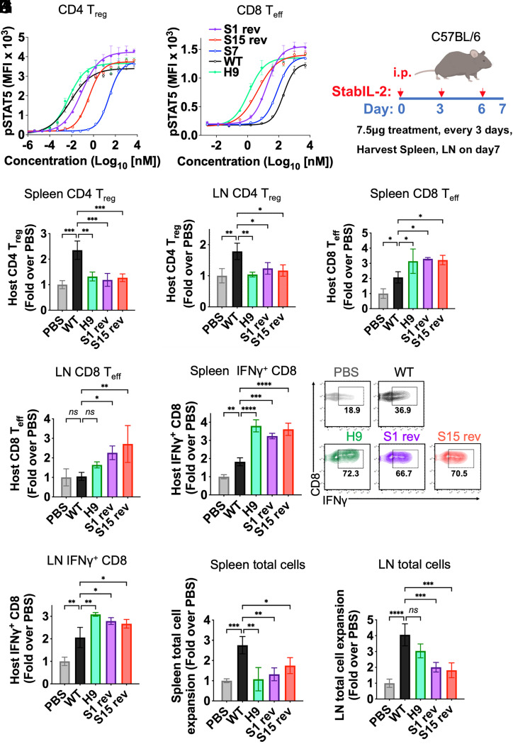
Fig. 4.
In vivo activity of stabIL-2 designs. (A and B) CD4+ Treg and CD8+ Teff were isolated from Foxp3-GFP reporter mice (C57BL6/J). A total of 250,000 cells were incubated with stabIL-2s for 15 min at 37 °C. Dose-response curve showing STAT5 phosphorylation in (A) CD4+ Tregs and (B) CD8+ cells following a 15-min cytokine stimulation. (C) Schematic of the in vivo immune cell profiling of stabIL-2 designs in mice. (D–G) Quantification of stabIL-2 designs administration on BL6 mice (B and C) Treg (CD4+CD25+Foxp3+)and (D and E) CD8+ Teffs (CD44+CD62L−). (H and I), Quantification of cytokine administration on C57BL/6 mice CD8+ production of IFN-γ upon ex vivo restimulation with phorbol 12-myristate 13-acetate (PMA) and ionomycin in (F) spleen and (G) lymph nodes (LNs). (J and K) Total cell count of immune cells in the (H) spleen and (I) LNs after stabIL-2 administration. Bar graphs show mean ± SD and were analyzed by one-way ANOVA relative to WT IL-2. Multiple comparisons were corrected using Dunnett’s test. Data are representative of three independent experiments. *P < 0.05; **P < 0.01; ***P < 0.001; ****P < 0.0001. ns, not significant.

