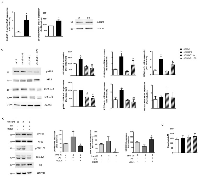
Figure 2.
SUCNR1 mediates basal and LPS-stimulated inflammatory pathways in intestinal epithelial cells. (a) HT29 cells treated with LPS 0.1 µg/mL during 24 h and graphs show mRNA and protein expression of SUCNR1 (n = 5). Image of a representative Western Blot of one independent experiment. (b) HT29 cells transiently transfected with a specific siRNA against SUCNR1 or ctrl and treated with LPS 0.1 µg/mL during 24 h. Graphs show protein expression of pERK-1/2, ERK-1/2, pNFкB and NFкB (n = 6) and mRNA expression of IL1B, iNOS, IL6 and TNF-a (n = 5). Image of a representative Western Blot of one independent experiment. (c) HT29 cells treated with vehicle or LPS (with or without the MEK inhibitor U0126 10 µM) over 2 h and graphs show protein expression of pERK-1/2, ERK-1/2, pNFкB, NFкB and IкB (n = 5). Image of a representative Western Blot of one independent experiment. (d) Graph shows succinate levels in supernatant of HT29 cells (n = 4). In all cases, bars in graphs represent mean ± SEM. * p < 0.05, ** p < 0.01 and *** p < 0.001 vs. vehicle cells. # p < 0.05 and ## p < 0.01 vs. the respective siCtrl cells. $ p < 0.05 vs. U0126 nontreated cells.

