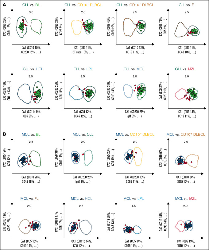
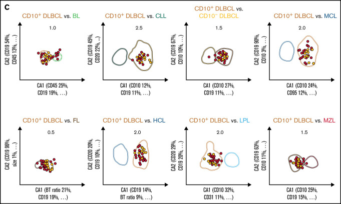
Figure 2.
Expert-independent classification of the CLL, MCL, and CD10+ DLBCL validation cohorts. Out of the total possible 36 2-dimensional CCA-based projections, the 8 projections that include CLL (A), MCL (B), and CD10+ DLBCL (C) are shown only. The X- and Y-axes of each plot represent CA1 and CA2. CA1 is the projection that captures most of the information for maximum separation between 2 B-CLPD entities; CA2 is the projection that provides the second-greatest amount of independent information for separation. Numbers in the top part of each plot represent the x fold SD of the immunophenotype shown. Numbers in brackets denote the relative contribution of markers to CA1 and CA2, respectively (see supplemental Table 4 for a full list of markers and coefficients). Each dot represents the median of 1 case from the validation cohort. (A) Cases included into all 8 representations for CLL are shown in green (n = 112); cases not included into all 8 plots for this leukemia are shown in red (“not classified”, n = 13). These 13 cases did not meet all 8 decision criteria for any other lymphoma. (B) Cases included into all 8 representations for MCL are shown in blue (n = 41); cases not included into all 8 plots for that lymphoma are shown in red (n = 14). Thereof, 13 cases did not meet all 8 decision criteria for any other lymphoma, and 1 MCL was misclassified as LPL (data not shown).


