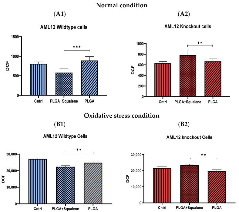
Figure 4.
Assessment of ROS production in normal mouse AML12 cells cell line (AML12 wild-type (WT)) and TXNDC5-deficient AML12 cells (AML12 knockout (KO)). (A,B) After treatment of cells with 30 µM of squalene NPs for 72 h, (A) ROS was measured in normal conditions, (B) oxidative stress circumstance by 25 mM of H2O2 for 3 h. (A1) potent reduction of ROS in squalene group in WT cells, and (A2) significant enhancement in KO cells were observed. (B1) Considerable decrement of ROS in squalene group in WT cells and (B2) remarkable increase in KO cells are indicated. Statistical analyses were done according to Mann–Whitney’s U-test; ** p< 0.01, *** p< 0.001.

