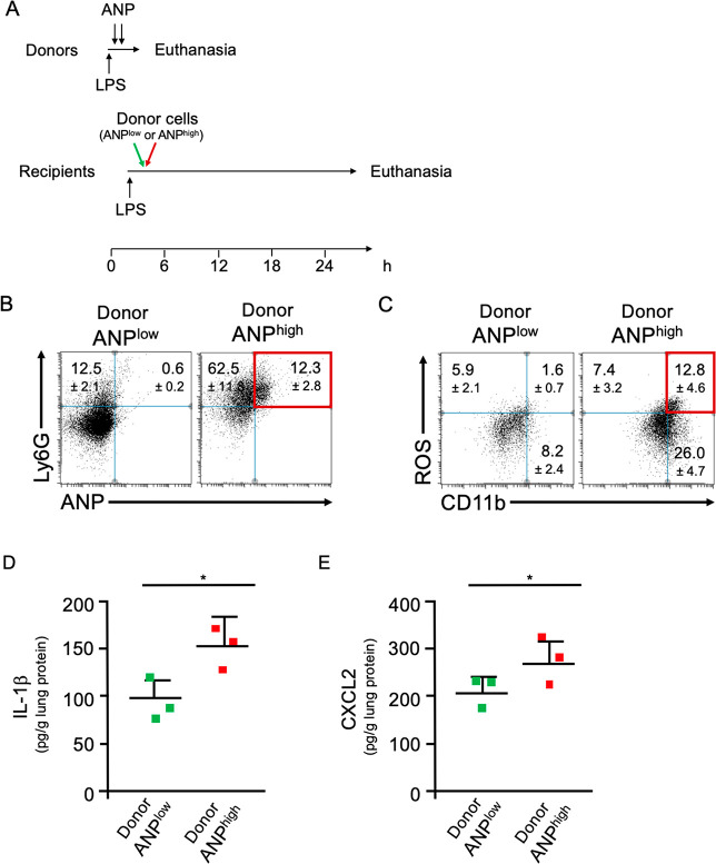
Figure 5.
ANPhigh PMN transfer inflammation. (A) Timeline of adoptive transfer. Donor mice were challenged with a lethal dose of LPS and injected with two doses of ANP labeled with the stable fluorochrome AF647. ANPhigh PMN (8 × 10⁵) or, as controls, an equal number of ANPlow lung Ly6G+ PMN were adoptively transferred by i.v. injection into syngeneic recipient mice. (B) Flow cytometric analysis of lung cells from mice that received ANPlow or ANPhigh donor cells. Dot blot. Percentages of Ly6G+ANP+ cells (red) were significantly greater in mice that received ANPhigh donor cells as compared to mice that received ANPlow donor cells. p < 0.001 (Student’s t test). (C) Flow cytometric analysis of lung cells from mice that received ANPlow or ANPhigh donor cells. Dot blot. Percentages of ROS+CD11b+ (red) were significantly greater in mice that received ANPhigh donor cells as compared to mice that received ANPlow donor cells. (D) Concentrations of IL-1β in lung tissue extracts from mice that have received ANPlow or ANPhigh donor cells. (E) Concentrations of CXCL2 in lung tissue extracts from mice that have received ANPlow or ANPhigh donor cells. Squares represent values from individual mice and lines indicate mean values + SD *p < 0.05 (Student’s t test). Representative data from 3 independent experiments are shown.

