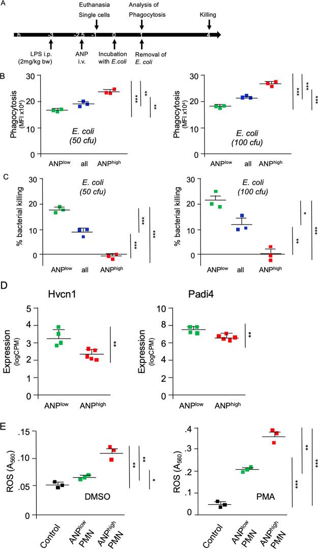
Figure 6.
Functional heterogeneity of lung PMN. (A) Timeline of assays. (B) E. coli phagocytosis. PMN were incubated with E. coli bacteria (50 cfu or 100 cfu) for 1 h. E. coli-specific fluorescence is shown. (C) Killing of intracellular E. coli bacteria. Single cell suspensions of lung unsorted Ly6G+ PMN (blue) or sorted according to endocytosis of ANP (low, green; or high, red). PMN were incubated with E. coli bacteria (50 cfu or 100 cfu) for 1 h, and then washed and incubated for additional 3 h to evaluate bacterial killing. E. coli-specific fluorescence at 4 h relative to E. coli-specific fluorescence at 1 h, corresponding to bacterial killing. Average (n = 3) of fluorescence detected at 1 h = 100%; % killing = 100 – percentage of fluorescence detected at 4 h post start of incubation. Markers represent results from individual mice*p < 0.005, **p < 0.002, ***p < 0.0002. (D) mRNA expression of hydrogen voltage gated channel 1 (Hvcn1) and peptidyl arginine deiminase 4 (Padi4) in ANPlow and ANPhigh lung PMN after LPS-challenge. (E) ROS production by ANPhigh is not impaired. Mice were injected i.p. with LPS and 2 h 30 min later with ANP (i.v.) and euthanized 30 min thereafter. Single cell suspensions of the lungs Ly6G+ PMN were sorted according to endocytosis of ANP. Cells were incubated and stimulated with DMSO or phorbol ester PMA. Control, unsorted PMN from naive mice. Squares represent results from individual mice. *p < 0.005, **p < 0.002, ***p < 0.0001.

