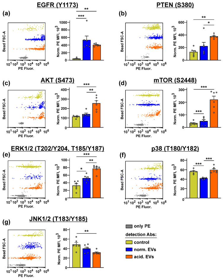
Figure 5.
Influence of “normal” and “acidified” EVs on the activity of the different intracellular signaling pathways in the keratinocytes. Het1-A cells were incubated with “normal” and “acidified” EVs for 48 h, and the phosphorylation of EGFR (Y1173) (a), PTEN (S380) (b), AKT (S473) (c), mTOR (S2448) (d), ERK1/2 (T202/Y204, T185/Y187) (e), p38 MAP kinase (T180/Y182) (f), and JNK1/2 (T183/Y185) (g) was assayed by the Bio-Plex magnetic beads assay. Representative distributions of the magnetic beads incubated with the lysates of the untreated (control) or treated by EVs’ keratinocytes and the beads stained only by PE are shown on the left panels (every bead probe was analyzed separately and combined on the one panel for illustration). Data on the analysis of the phosphorylation level of the messengers are shown on the right panels. Data were acquired by the Attune NxT flow cytometer and presented as normalized MFI ± SEM (n = 6). * (p < 0.05), ** (p < 0.01), and *** (p < 0.001) indicate significant difference between the data groups by the one-way ANOVA followed by Tukey’s post hoc test.

