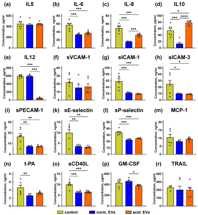
Figure 7.
Effect of “normal” and “acidified” EVs on secretion of the different cytokines and adhesion factors by the keratinocytes. Het1-A cells were incubated with “normal” and “acidified” EVs for 48 h, and the concentration of IL5 (a), IL10 (d), IL12 (e), GM-CSF (p), and TRAIL (r) was assayed by ELISA. Concentration of IL6 (b), IL8 (c), sVCAM-1 (f), sICAM-1 (g), sICAM-3 (h), sPECAM-1 (i), sE-selectin (k), sP-selectin (l), MCP-1 (m), t-PA (n), and sCD40L (o) was analyzed by the Flow Cytomix kits. Data presented as the normalized protein concentration ± SEM (n = 6). Control corresponds to the untreated cells. * (p < 0.05), ** (p < 0.01), *** (p < 0.001), and **** (p < 0.0001) indicate significant difference between the data groups by the one-way ANOVA followed by Tukey’s post hoc test.

