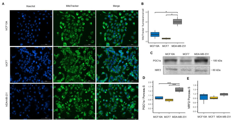
Figure 2.
Increased mitochondrial mass in MDA-MB-231 cell line. (A) Confocal microscopy images of MitoTracker Green fluorescence to assess mitochondrial mass in MCF10A, MCF7 and MDA-MB-231 cells, and total nuclei were stained with Hoechst (blue). (B) Mitochondrial mass levels were quantified as MitoTracker green fluorescence intensity/cell ratio. Data were obtained from three biological replicates. The data are presented as mean ± SD of cells (n = 151–251). * p < 0.05. (C) Representative blots. (D,E) Expression of mitochondrial biogenesis markers: peroxisome proliferator-activated receptor-gamma coactivator-1alpha (PGC-1α), n = 6 and nuclear respiratory factor 2 (NRF2), n = 3. Densitometry values were normalized by Ponceau S red staining. The data are presented as mean ± SD. *** p < 0.001.

