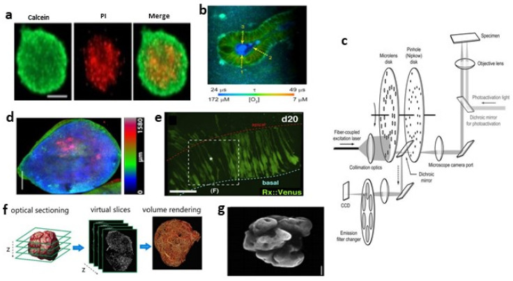
Figure 2.
(a) WFFM was used to observe viable and dead cells in the cancer spheroid. Stained with calcein-AM (green) and propidium iodide (PI, red). Reproduced from Reference [81] under the Creative Commons License (CC BY 4.0); (b) FLIM imaging revealed presence of O2 micro-gradients between basal and apical membranes in resting organoids. Reprinted from Biomaterials, 146, D.B.; Okkelman, I.A.; Foley, T.; Papkovsky Dmitriev R.I., Live cell imaging of mouse intestinal organoids reveals heterogeneity in their oxygenation, 86–96,Copyright (2022), with permission from Elsevier; (c) Schematic drawing of SDC microscopy. Reprinted from Methods in Enzymology, 504, Stehbens, S.; Pemble, H.; Murrow, L.; Wittmann, T., Imaging intracellular protein dynamics by spinning disk confocal microscopy, 293–313, Copyright (2022), with permission from Elsevier; (d) Color-coded SDC image of endogenous GFP in a human cerebral organoid. Adapted from [84]. (e) Multiphoton images showing interkinetic nuclear migration of retinal progenitors in the day-20 hESC-derived optic vesicle epithelium. Reprinted from Cell Stem Cell, 10, Nakano, T.; Ando, S.; Takata, N.; Kawada, M.; Muguruma, K.; Sekiguchi, K.; Saito, K.; Yonemura, S.; Eiraku, M.; Sasai, Y., Self-formation of optic cups and storable stratified neural retina from human ESCs, 771–785, Copyright (2022), with permission from Elsevier; (f) Schematic drawing of light sheet 3D reconstruction. Reprinted from Neoplasia, 16, Dobosz, M.; Ntziachristos, V.; Scheuer, W.; Strobel, S. Multispectral fluorescence ultramicroscopy: Three-dimensional visualization and automatic quantification of tumor morphology, drug penetration, and antiangiogenic treatment response, 1–13, Copyright (2022), with permission from Elsevier; (g) Light sheet image of a 6-week-old human cerebral organoid. Reprinted from Cell Stem Cell, 20, Li, Y.; Muffat, J.; Omer, A.; Bosch, I.; Lancaster, M.A.; Sur, M.; Gehrke, L.; Knoblich, J.A.; Jaenisch, R. Induction of Expansion and Folding in Human Cerebral Organoids, 385–396, Copyright (2022), with permission from Elsevier.

