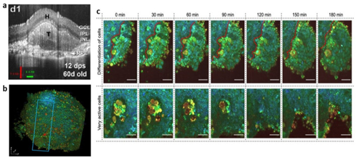
Figure 4.
(a) In vivo development of retinal organoid transplant monitored by OCT. Reproduced from Reference [171] under the Creative Commons License (CC BY 4.0); (b,c) D-FFOCT 3D image (b) differentiation process is shown in the top row and the cell’s dynamic active region is shown in the bottom row (c) of hiPSC-derived retinal organoids. Reproduced from Reference [173] under the Creative Commons License (CC BY 4.0).

