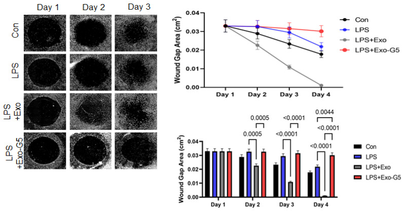
Figure 6.
HDF cells were grown on a collagen scaffold to mimic 3D wound healing. Chronic inflammation was induced for 4 days using 5 ng/mL LPS for 6 h followed by treatment with hASC exosomes (Exo) or exosomes depleted of GAS5 (Exo-G5) for 18 h. LPS was maintained in the media along with the treatment. The 3D wound model was maintained at 37 °C and wound closure was imaged every 24 h using a Keyence BX810 microscope (n = 3). Wound closure was calculated each day using a Keyence’s Cell migration assay with Hybrid cell count software to calculate wound gap area. Statistical analysis was performed by one-way ANOVA and significant p-values (<0.05) are indicated on the graph.

