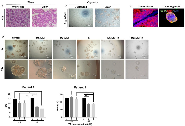
Figure 6.
TQ radiosensitizes patient 1-derived rectal cancer organoids and reduces their organoid-forming ability and size. (a) Representative images of H&E stain of unaffected rectum and rectal cancer tissue from patient 1. (b) Representative bright-field images of organoids derived from unaffected rectum and rectal cancer samples (patient 1). (c) Immunofluorescent images of rectal tumor issues and organoids stained for CD44 and CK19. Images were obtained using confocal microscopy. (d) Representative bright-field images of organoids derived from rectal cancer patient 1 sample and treated with TQ (3 and 5 µM), radiation (2 Gy), or combinations. Fresh unaffected and tumor tissues were digested, and single cells were resuspended in 90% Growth Factor reduced Matrigel and 10% serum-free colon media and allowed to grow in serum-free colon media (without treatment). Generated organoids are referred to as G1 organoids. Organoids were propagated to G2 and treated with TQ, IR, or combinations. OFC and size were calculated, and average values were reported as mean ± SEM (* p < 0.05, ** p < 0.01, *** p < 0.001). Images were visualized by Axiovert inverted microscope at 10× magnification. Scale bar for bright-field images is 100 µm and for immunofluorescent images is 50 µm.

