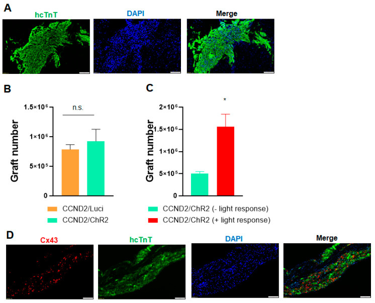
Figure 6.
Immunohistological analysis of engrafted hiPSC-CMs. (A) Heart sections from light-responsive mice that had been sacrificed 6 months after myocardial infarction induction and treatment with PBS and hiPSC-CMs were stained for the presence of hcTnT. Nuclei were counterstained with DAPI. Scale bar = 100µm. (B,C) The engraftment rate was calculated by dividing the number of cells that expressed hcTnT by the total number of cells administered and expressed as a percentage. *—p < 0.05 versus mice receiving hiPSC-CCND2OE/ChR2OECMs but not responsive to light; Student’s t-test. n = 10 mice in hiPSC-CCND2OE/LuciOECMs group; n = 10 mice in hiPSC-CCND2OE/ChR2OECMs group (n = 6 not responsive to light and n = 4 responsive to light). n.s. indicates no significant difference. (D) Engrafted hiPSC-CCND2OE/ChR2OECMs were identified in mouse hearts at 6 months after myocardial infarction induction and cell transplantation via immunofluorescent staining for the expression of human cTnT. Both transplanted cardiomyocytes and native cardiomyocytes were stained with antibodies against Connexin-43. Images were from heart sections of light-responsive mice. Scale bar = 100 µm.

