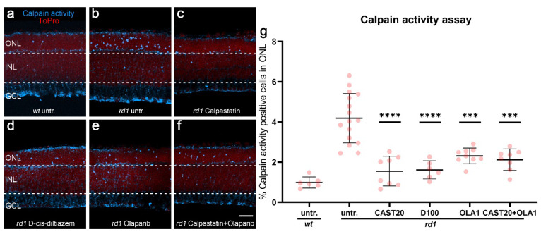
Figure 1.
Effects of calpastatin, D-cis-diltiazem, Olaparib, and combination treatments on calpain activity. The calpain activity assay (blue) was performed on unfixed wt (a) and rd1 retinal cross-sections. ToPro (red) was used as nuclear counterstaining. Untreated rd1 retina (untr.; b) was compared to retina treated with either calpastatin (c), D-cis-diltiazem (d), Olaparib (e), or calpastatin and Olaparib combined (f). The scatter plots show the percentages of outer nuclear layer (ONL) cells positive for calpain activity (g) in wt and rd1 retina. Statistical significance was assessed using one-way ANOVA and Tukey’s multiple comparison post hoc testing performed between the control (rd1 untreated) and 20-μM calpastatin (CAST20), 100-μM D-cis-diltiazem (D100), 1-μM Olaparib (OLA1), and 20-μM calpastatin combined with 1-μM Olaparib (CAST20+OLA1). All treatments reduced the calpain activity in rd1 ONL; however, there was no added synergistic benefit from the CAST20+OLA1 combination. Untr. wt: 6 explants from 3 different mice; untr. rd1: 16/16; CAST20 rd1: 8/8; D100 rd1: 6/6; OLA1 rd1: 9/9; CAST20+OLA1 rd1: 8/8; error bars represent SD; *** = p ≤ 0.001 and **** = p ≤ 0.0001. INL = inner nuclear layer, GCL = ganglion cell layer. Scale bar = 50 µm.

