Scheme A2.
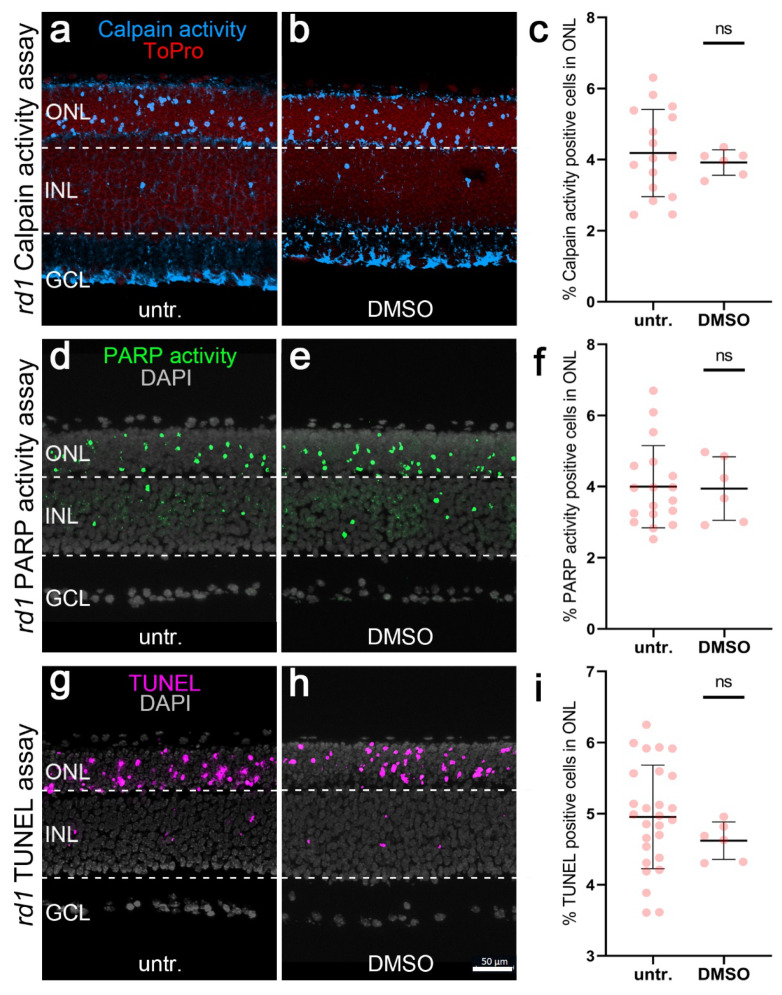
Effect of DMSO on calpain activity, PARP activity, and TUNEL staining in rd1 retinal explants. Calpain activity (blue), PARP activity (green), and TUNEL (magenta) were used in rd1 retinal explant cultures. ToPro (red) and DAPI (grey) were used as nuclear counterstains. rd1 control retina (untr.; a,d,g) was compared to retina treated with 0.1% DMSO (DMSO; b,e,h). There was no difference between positive cells detected in the rd1 outer nuclear layer (ONL), with or without DMSO. The scatter plot (e) shows the percentage of calpain activity (c), PARP activity (f), and TUNEL-positive cells (i). In the calpain activity assay, untr. rd1: 16 explants derived from 16 different animals; DMSO rd1: 6/6. In the PARP activity assay, untr. rd1: 18/18; DMSO rd1: 6/6. In the TUNEL assay, untr. rd1: 27/27; DMSO rd1: 6/6; error bars represent SD; ns = p > 0.05, INL = inner nuclear layer, GCL = ganglion cell layer. Scale bar = 50 µm.

