Scheme A3.
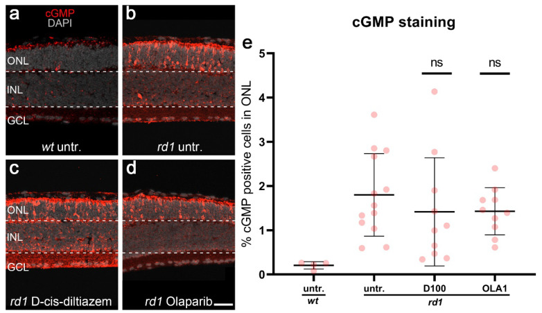
Photoreceptor accumulation of cGMP in different experimental conditions. cGMP immunostaining (red) was used in wt and rd1 retinal explant cultures. DAPI (grey) was used as a nuclear counterstain. In wt retina (untr.; a), only a few cells in the outer nuclear layer (ONL) were labeled cGMP-positive. rd1 control retina (untr.; b) was compared to retina treated with either 100-µM D-cis-diltiazem (D100, c) or 1-µM Olaparib (OLA1, d). Statistical significance was assessed using one-way ANOVA, and Tukey’s multiple comparison post hoc testing was performed between control, 100-μM D-cis-diltiazem (D100), and 1-μM Olaparib (OLA1). Note the large numbers of cGMP expressed in the rd1 ONL. The scatter plot (e) shows the percentage of cGMP-positive cells in the ONL. Neither D-cis-diltiazem or Olaparib reduced the cGMP-positive cells. Untr. wt: 4 explants from 2 different mice; untr. rd1: 13/13; D100 rd1: 10/10; OLA1 rd1: 10/10; error bars represent SD; ns = p > 0.05. INL = inner nuclear layer, GCL = ganglion cell layer. Scale bar = 50 µm.

