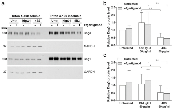
Figure 8.
4B3 antibody-induced Dsg3 depletion occurs from the non-desmosomal and desmosomal pools and cannot be rescued by efgartigimod. hTert cells were plated on 6-well plates, grown in KGM2 medium with 0.05 mM CaCl2 until confluent, and then switched to KGM2 with 2 mM CaCl2 for 24 h. Thereafter, the cells were either left untreated, or efgartigimod (25 µg/mL) was applied for 30 min prior to a 24 h treatment with 4B3 (50 µg/mL) antibodies. As controls, mock incubation (untreated) and an isotype-matched human IgG1 (50 µg/mL) were included. Sequential detergent extraction was performed, resulting in Triton-soluble (non-desmosomal) and Triton-insoluble (desmosomal) pools of proteins. (a) Western blot analysis of the fractions was performed to detect Dsg3 and Dsg1. Analysis of GAPDH level was included to ensure equal loading and the purity of the detergent insoluble fraction. Equal percentage of each fraction was loaded. Western blot signals for Dsg3 in the non-desmosomal (b) and desmosomal (c) pools were quantified using ImageJ software, normalized against GAPDH levels, and expressed as relative amounts compared to the untreated controls. The error bars represent the SD of four independent experiments. Statistical analysis was done using two-way ANOVA with Bonferroni’s post-test. Statistically significant differences are indicated by * = p ≤ 0.05, ** = p < 0.01.

