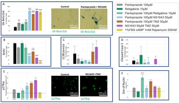
Figure 14.
Senescence and Proliferation Analysis of U87 Cells Reveals that Treatments with Pantoprazole or NS164 with TMZ Increased Senescence, Decreased BrdU Incorporation, and Increased a p27Kip1 after 6 days A senescence associated beta-galactosidase stain was conducted and scored by eye. Immunofluorescence of cells was conducted and analyzed with CellProfiler for integrated fluorescence intensity or presence or absence of a cellular signal. (A) Stain of senescence associated beta-galactosidase stain (SA-Beta Gal). (B) Stain of bromodeoxyuridine incorporation (BrdU). (C) Stain of the microtubule-associated protein light chain 3 B (LC3B). (D) Stain of cleaved caspase 3 (Casp 3). (E) Stain of cyclin-dependent kinase inhibitor 1B (p27Kip). (F) Size of Nuclei, determined by area of the Hoechst stain. Treatments corresponding to the colored bars are outlined in the figure itself. The log of the fold change in intensity was compared between single treatments and combined treatments, except the positive control, with significant values shown in the color of the treatment compared. The initial fluorescence intensities were compared to their corresponding control, with significant values shown under the bars. The logit of the percent positive cells was compared between single treatments and control, in cases of 0 values, the arcsine transformation was used. Significance was expressed as, ***: p < 0.001, **: p < 0.01, *: p < 0.05 (one-way ANOVA with Tukey post hoc analysis n > 3 technical replicates).

