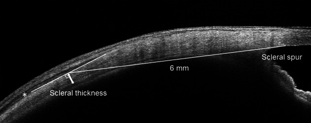
Fig. 1.

Scleral thickness measurement using anterior-segment optical coherence tomography. The anterior scleral line was determined based on the low reflective bands of the rectus muscles (asterisk). The posterior scleral line was determined by the difference in reflection intensity between the choroid and sclera. Scleral thickness measurements (double-headed arrow) were conducted vertically at the point 6 mm posterior to the scleral spur in the superior, temporal, inferior, and nasal directions.
