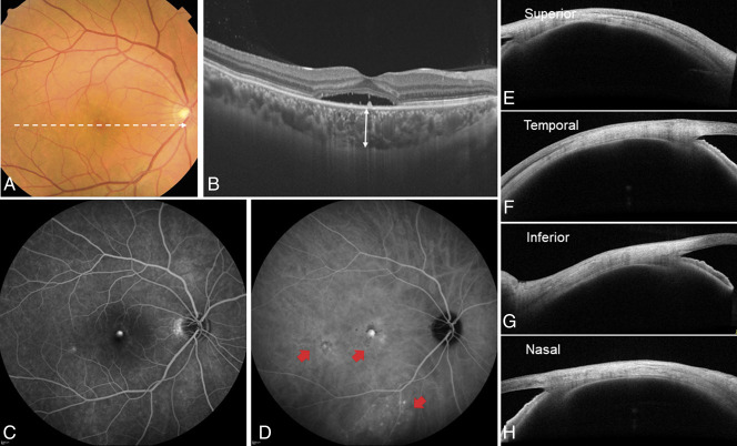
Fig. 3.
A representative case of the right eye of a 43-year-old man with central serous chorioretinopathy without ciliochoroidal effusion. The spherical equivalent refractive error was +1.125 diopters, and the axial length was 23.21 mm. A. Color fundus photograph showed a SRD in the macular area. B. Optical coherence tomography along the white dotted line in the color fundus photograph revealed SRD and pachychoroid with dilated choroidal vessels. The subfoveal choroidal thickness (double-headed arrow) was 360 µm. C. Fluorescein angiography demonstrated typical leakage in the central macula. D. Indocyanine green angiography revealed multifocal areas of CVH (red arrows). The total area of CVH was 7.22 mm2. E–H. Anterior-segment OCT demonstrates cross-sectional images of the anterior sclera in four directions. The scleral thickness at the superior, temporal, inferior, and nasal directions was 403 µm, 370 µm, 415 µm, and 405 µm, respectively.

