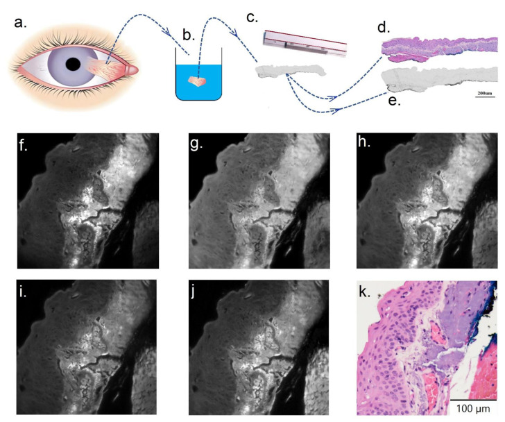
Figure 2.
(a–e) Sample preparation and histological assessment. (a) Ocular surface biopsy collected from patients. (b) Histology sample processed following formalin fixation into paraffin embedded sections. (c) Two adjacent sections were cut using a microtome and then dewaxed. (d) Example cut tissue section, which was H&E stained and coverslipped for histology assessment and used as reference. (e) The unstained tissue section adjacent to that shown in (d). Such sections were placed on a slide, coverslipped, and used for multispectral imaging analysis. (e–j) Example tissue images in selected channels (channels number 3, 16, 22, 31, and 45, respectively). (k) H&E stained section of example tissue shown in (e–j).

