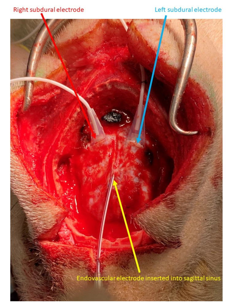
Figure 1.
Endovascular and subdural electroencephalograms. Four-contact subdural electrodes were placed in each hemisphere through a linear dural incision (blue and red arrows). The endovascular electrode was inserted into the sagittal sinus through the outer needle of the venous catheter needle (yellow arrow).

