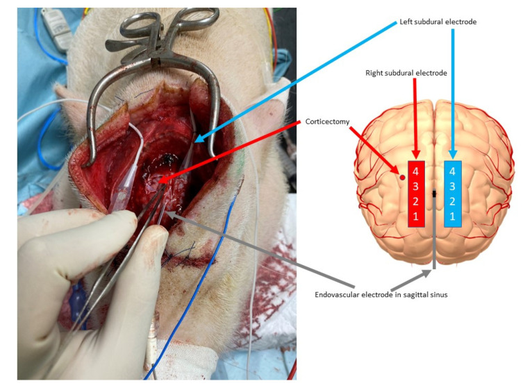
Figure 3.
Corticectomy for placement of the epileptogenic focus and positioning of the subdural and endovascular electrodes. On the right frontal area, corticectomy for the epileptogenic focus was performed, 5 mm lateral to the epileptogenic focus and between contacts 3 and 4 of the right subdural electrode (red arrow). The left subdural electrode (sky blue arrow) and endovascular electrode (gray arrow) were already placed (left). The location of each electrode and the artificially created epileptogenic area are shown schematically (right).

