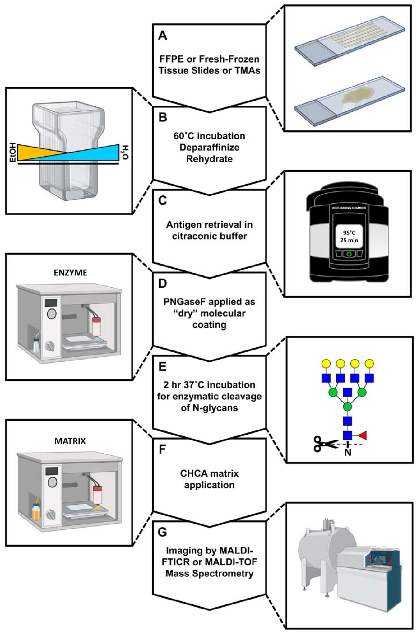
Figure 4. A standardized workflow for in-situ N-glycosylation analysis by MALDI-IMS.

A) FFPE or FF tissue slides. B) Dewaxing and rehydration of tissue specimens prepares them for antigen retrieval and removes signal-suppressing lipids and metabolites. C) Heat-induced epitope exposure in low-pH citraconic buffer. D) Use of an automated solvent sprayer to apply a dry molecular coating of peptide N-glycosidase F. E) Enzymatic cleavage of N-glycans from their glycoprotein carriers. F) Application of a crystalline organic acid matrix by an automated solvent sprayer as in D. G) Spatial analysis of released N-glycans by matrix-assisted laser desorption/ionization and FT-ICR MS detection.
